Microfluidics and microelectronics are two technologies that are quite related, in fact, conventional integrated circuit manufacturing techniques (microelectronics) as photolithography are the same as those used when manufacturing microfluidic devices but with some variations (soft lithography and rapid soft lithography).
For its part, microfluidics is being used for the development of increasingly complex medical devices and that is where microelectronics intervene to increase the functionality of such devices, In this opportunity I will tell you about the proposal of UTEC for the development of a diagnostic device of malaria that integrates these two technologies.
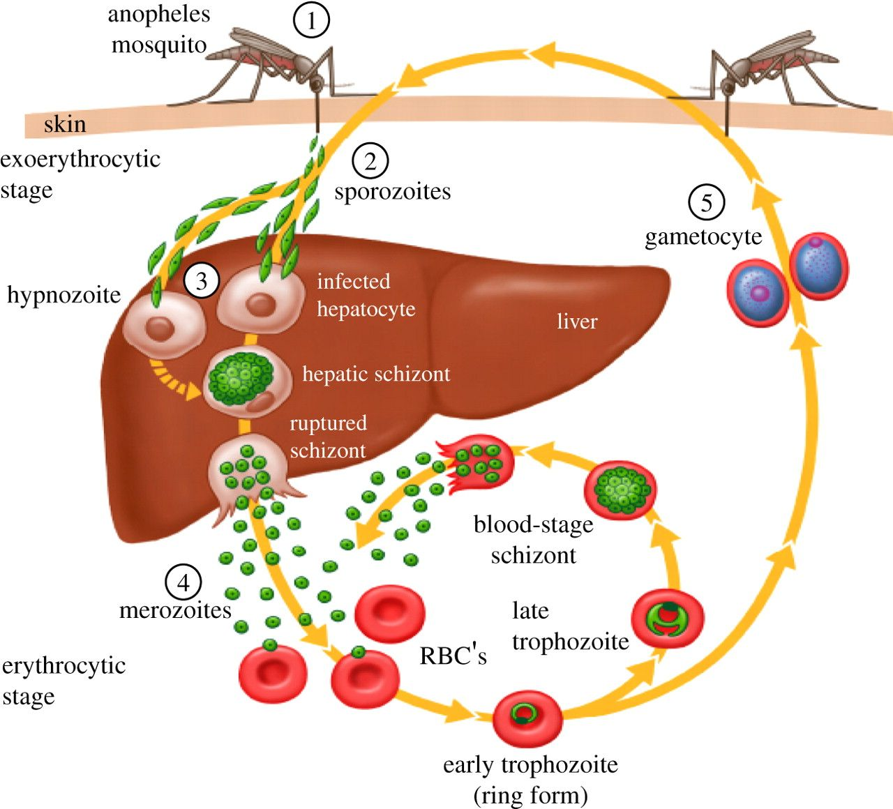
Figure 1. Plasmodium spp. (REF: https://es.scribd.com/document/443591846/Pathogenesis-of-malaria-UpToDate)
Malaria is a disease caused by parasites of the genus Plasmodium and transmitted by Anopheles mosquitoes, when these parasites infect red blood cells generate changes in the mechanical and electrical properties of erythrocytes, for example, they may become stiffer or softer than a healthy cell and electrical impedance has been observed to be higher in cells infected with plasmodium falciparum. Figure 1 illustrates the life cycle of the parasite.
Understanding the aforementioned changes caused by malaria in erythrocytes in UTEC, a modular microfluidic system composed of two main parts was proposed: an inertial separation module and a microfluidic bioimpedance module. Figure 2 shows the modular system.
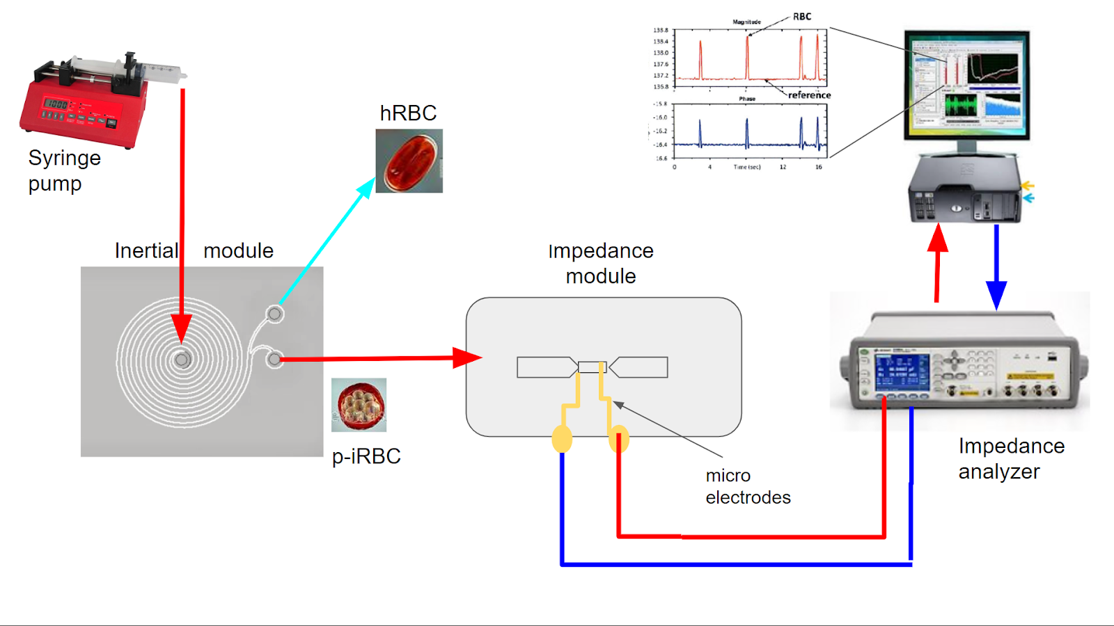
Figure 2: Modular microfluidic system for malaria diagnosis
As we can see in Figure 2, in the proposed system, the sample first enters the inertial separation module, this module takes advantage of changes in erythrocyte stiffness to, through microhydrodynamic interactions, focus cells in positions other than the spiral-shaped microchannel so that they can separate, healthy from infected, at the final fork. This stage serves as a pre-concentrator of red blood cells that have been invaded by the parasite.
The infected cells pass to the next module, which will measure the impedance of each individual cell to determine/confirm its health status and thus make a better diagnosis. It is at this stage where microelectronics intervene since the impedance module must not only have a microchannel but two microelectrodes located at the bottom of the channel with which the electrical measurements will be made, These microelectrodes are 20 micrometers wide, 20 micrometers apart and only 100 nanometers thick. Figure 3 shows the modules already manufactured (1, 4), a microscope image of the microelectrodes (2) and a diagram thereof (3).
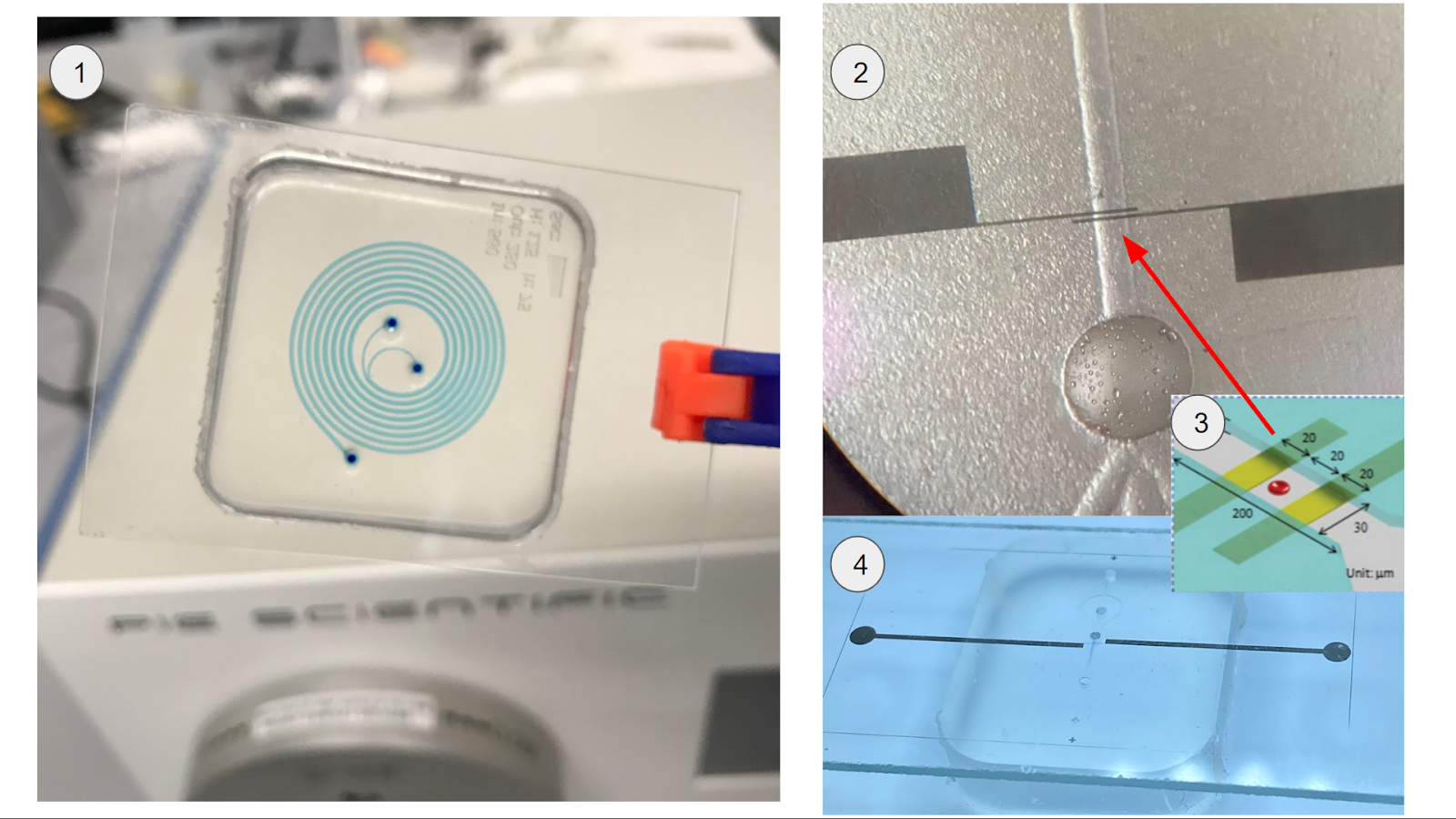
Figure 3. (1) Inertial separation spiral, (2) microscope image of microelectrodes passing through the microchannel. (3) microelectrodes and microchannel scheme, (4) Manufactured microfluidic impedance module.
Finally, using an instrument called an impedance analyzer, a voltage signal is injected and the generated current is read to calculate the impedance in a given frequency range. The project is in progress with interesting results that will soon be published in scientific papers.
You may be wondering how have we managed to manufacture these devices in UTEC? , this I will tell you in detail in a next installment, for now I will just tell you that we had to use a lot of technology and creativity.