Decellularization is the process by which all cellular and nuclear components of a tissue or organ are removed to prevent an initial immune response while preserving the ultrastructure and composition of the native extracellular matrix (ECM), because the morphological and biochemical conditions of the elularized matrices are favorable for adhesion, proliferation and cell differentiation. Decellularized structures also show a decreased immunogenic response in the host after implantation compared to allogeneic and xenogenic transplants because immunogenicity is linked to intracellular components that are eliminated during the decellularization process.
Decellularized animal tissues have long been used in tissue engineering, in the form of porous solids, powders and gels to formulate hydrogels, yielding promising results, Despite this, animal and human sources are controversial and are affected by limited availability, high production costs and ethical concerns. Moreover, variability between different donors is a critical issue in clinical applications. De-elularized plant tissue scaffolding could overcome availability problems, high costs and ethical concerns related to the use of animal sources, given its easy availability, low cost, ease of use and absence of ethical issues, Moreover, plant tissues exhibit good cytocompatibility and biocompatibility.
In the plant kingdom there is a wide variety of architectures; therefore, scaffolding derived from decellular plants could be selected depending on their structure and native properties to mimic a multiplicity of mammalian tissues.
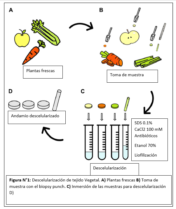
In this paper the authors chose, apple, carrot and celery to decellularize them because they are characterized by various internal architectures, characterizing them morphologically, They physically and mechanically investigated the potential in vitro as scaffolding for regeneration in adipose, bone and tendon tissue.
To test the decellularization of the vegetable tissues, first cut into slices with 2 mm thickness, with a biopsy punch cylindrical samples of 10 mm thickness were extracted, for mechanical tests samples of 4 mm diameter and length L = 25 mm were extracted.
To decellularize the plant tissue, they immersed each sample in 5 ml of 0,1 % w/v sodium dodecylsulphate solution (SDS) for 48 h at room temperature with continuous agitation. After 24 h, the samples were probed for 5 min at 40 °C and the SDS solution was changed, the samples were washed three times in distilled water and in CaCl2 100 mM for 24 h.
Samples of plants (APctr, CActr and CEctr) that were not treated by the decellularization process were used as control. Both the decellularized and control samples were washed three times in distilled water and then incubated with 1 % w/v penicillin/streptomycin and 1 % w/v amphotericin B for 3 h with agitation at 140 rpm. Finally, the samples were disinfected in 70% v/v ethanol solution for 1 h, washed three times in sterile distilled water and frozen at -20 C overnight. The structures obtained were then freeze-dried and sterilized by UV radiation for 15 min on each side.
After the decellularization procedure, volumetric changes and structural damage were evaluated in the different samples, finding that the maximum contraction recorded in the scaffolding was 6% (carrot).
Each sample absorbed water (24 to 66 times its initial anhydrous weight) and reached a plateau value during the first week of immersion, demonstrating the ability of the samples to retain absorbed fluid. Apples reached stable weights after 24 h and carrot and celery samples after 7 days of incubation. The weight variation for apple and carrot derived samples was 4500 and 2450 %, respectively, without differences between the decellularized tissues and the native ones (p > 0.05). The weight variation in the celery scaffolding was 6660 %, with a significant increase in water absorption for the elularized samples compared to native tissue (P < .).
Thus proving the ability to conserve water after decellularization. With electron microscopy they observed
These data prove that the peculiar presence of a large amount of water in plant tissues is preserved after decellularization and that the structure obtained is thus able to absorb fluids, essential for the survival of cells in tissue volume. In fact, water retention is a crucial aspect for the development of successful scaffolding capable of replacing natural body tissues. Then a scanning electron microscopy analysis was performed where they observed that the decellularized plant tissues preserved a highly porous three-dimensional structure, without alteration of the morphology. Cell cytotoxicity tests found 95% cell viability and no significant differences in cell growth in the control and decellularization groups.

Apple-derived scaffolding had a relatively large and homogeneous porosity, suitable for the regeneration of adipose tissue, with a pore size greater than 100 µm is suitable for an efficient supply of oxygen and nutrients. Elularized and hydrated apple-derived samples were characterized by a compression module of 4.17 0.17 kPa; this is comparable to native human adipose tissue. No statistical differences were observed between the elastic modules of the elularized and control samples. Using the preadipogenic cell line (3T3-L1) the metabolic activity of the cells grown in apple-derived scaffolding increased over the 14 days of cultivation was measured, demonstrating the ability of apple scaffolding to sustain the growth and proliferation of preadipocytes. After 14 days of culture with the Oil Red O stain differentiated adipose cells were observed, which possessed an intracellular accumulation of lipids
The decallularized carrot scaffolding presented a homogeneous structure with larger pores in the peripheral region (130 26 µm average size) compared to the pores located in the central region (70 12 µm average size), which are comparable to those used for bone tissue. Compatibility tests were performed in vitro using a preosteoblast cell line (MC3T3-E1). Metabolic activity was stable until 7 days after planting, carrot scaffolding maintained the growth and proliferation of preosteoblastic cells. The activity of ALP alkaline phosphatase was investigated after 14 days of cultivation in scaffolding derived from carrots. . The cells uniformly colonized the pore walls of carrot scaffolding, assuming an elongated morphology, typical of attached cells.
The scaffolding derived from celery was characterized by a longitudinally oriented structure, formed by pores of 125 11 µm aligned to form parallel channels, this type of structure mimics the alignment of tendons. These scaffolding withstood 20% stress without failure. Elastic modulus E of decellularized celery scaffolding is equal to 0.59 0.09 MPa, with no statistical difference (p < 0.05) with respect to control. Direct cytocompatibility in vitro was investigated using L929, a fibroblast cell line. Metabolic activity increased until a plateau value was reached. LIVE/DEAD staining confirmed the presence of viable L929 cells attached to the decellular celery scaffolding. Cellular alignment was observed in several regions of the celery surface by VIVO/DEAD staining, The samples had an alignment angle <20 This is comparable to the percentage of aligned cells observed in previously developed oriented scaffolding aimed at tendon tissue engineering. Randomly organized regions were also detected, where cells are characterized by a round morphology without preferential orientation, which is consistent with the structure of native tendons, characterized by aligned collagen fiber bundles surrounded by a soft interfascicular matrix, with a less defined and oriented structure.
References
[1] N. Contessi Negrini, N. Toffoletto, S. Farè, and L. Altomare, “Plant Tissues as 3D Natural Scaffolds for Adipose, Bone and Tendon Tissue Regeneration,” Front. Bioeng. Biotechnol., vol. 8, no. June, pp. 1-15, 2020, doi: 10.3389/fbioe.2020.00723.