Author: Luz Pérez – Tissue Engineering and Synthetic Biology Laboratory. Bioengineering Department
Cancer is a multifaceted pathology, in which tissue grows uncontrollably due to the altered expression and behavior of proliferative and survival signals, in which cellular and acellular elements interact to drive progression and, at worst cases, metastasis. Current methods (monolayer or 2D cell cultures and animal models) to investigate the heterogeneous nature of cancer are inadequate. Monolayer cell cultures insufficiently reflect the physiological response pattern of the in vivo situation due to fundamental geometric differences between two-dimensional (2D) cultures and three-dimensional (3D) solid tumors. It is necessary to generate a 3D architecture of tumor cells in vitro to simulate the multicellular microenvironment when investigating tumor cell physiology and response to therapeutic agents.
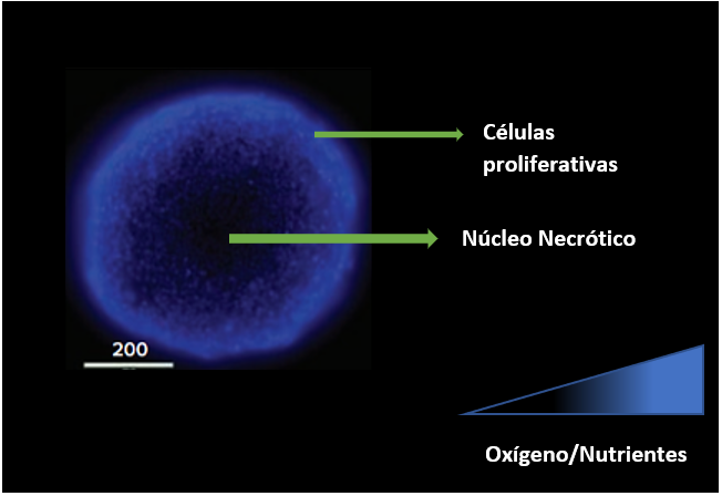
Spheroids are formed when cells are allowed to grow in suspension, as a result of which they aggregate, either on their own or with the help of extracellular matrices. The formation of spheroids is possible thanks to membrane proteins (integrins) and extracellular matrix proteins. These steps occur in spheroid formation: (i) dispersed cells aggregate due to ECM fibers containing RGD motifs that allow the cell surface integrin to bind and this leads to the expression of cadherins, (ii) cadherins accumulates on the surface of the cell membrane, (iii) the cadherin-cadherin bond between neighboring cells makes it possible to tighten the connections between cells and spheroids are formed; which are considered an improved in vitro model to mimic the biological properties of micrometastases and distal regions of tumor vessels because they retain the architecture and many morphological and physiological characteristics of their tumor counterparts. Multicellular tumor spheroids have been widely used as a 3D model to study the proliferation, apoptosis, differentiation, gene expression, and metabolism of tumor cells in a multicellular context since tumor cells in spheroids show a greater degree of morphological and functional differentiation than the cells grown in monolayer culture. Also showing growth kinetics, metabolic rates and resistance to radiotherapy and chemotherapy similar to tumor cells in vivo. Increased cell-cell and cell-matrix contacts in compact spheroids not only alter gene expression patterns and resistance to antineoplastic treatment, they also limit the diffusion of nutrients, oxygen, and waste products into and out of the spheroids, giving rise to a more layered composition, with the border of the spheroids consisting of proliferating cells, followed by a layer of quiescent cells in the middle and an inactive interior with necrotic cells in the nucleus of the sphere.
The quality of the spheroids can also be assessed using cell labeling techniques. For example, cell aggregates will stain homogeneously with a specific stain such as Masson’s trichrome stain (red color is associated with collagen deposition), Hematoxylin-eosin (blue, cell nuclei; pink some matrix proteins), toluidine blue (stains DNA) to or by different markers such as caspase 3 (apoptosis), EF5, hypoxia-inducible factor (hypoxia), Ki 67 (cell proliferation). Also on the market, there are fluorescent stains such as the live/dead cells kit, to be able to evaluate the number of live and dead cells of the spheroid.
Bibliographic references:
Białkowska, K., Komorowski, P., Bryszewska, M., & Miłowska, K. (2020). Spheroids as a type of three-dimensional cell cultures — examples of methods of preparation and the most important application. International Journal of Molecular Sciences, 21 (17), 1–17. https://doi.org/10.3390/ijms21176225
Bianchi, M. E., & Mezzapelle, R. (2020). The Chemokine Receptor CXCR4 in Cell Proliferation and Tissue Regeneration. Frontiers in Immunology, 11 (August), 1–8. https://doi.org/10.3389/fimmu.2020.02109
Costa, E. C., Moreira, A. F., de Melo-Diogo, D., Gaspar, V. M., Carvalho, M. P., & Correia, I. J. (2016). 3D tumor spheroids: an overview on the tools and techniques used for their analysis. Biotechnology Advances, 34 (8), 1427-1441. https://doi.org/10.1016/j.biotechadv.2016.11.002
Ivascu, A., & Kubbies, M. (2006). Rapid generation of single-tumor spheroids for high-throughput cell function and toxicity analysis. Journal of Biomolecular Screening, 11 (8), 922–932. https://doi.org/10.1177/1087057106292763
Molecular Devices Application Note: High-Throughput Confocal Imaging of Spheroids for Screening Cancer Therapeutics https://www.moleculardevices.com/en/assets/app-note/dd/img/high-throughput-confocal-imaging-of-spheroids- for-screening-cancer-therapeutics # gref